Thanks to the imaging techniques developed with today’s technologies, computed tomography (CT) can assess possible diseases of the cardiovascular system in seconds. It is a method to visualize the blood vessels of the cardiovascular system because it can take images up to 16 cm without moving the table when necessary. Hospitals and preventative health scanning facilities such as ViaScan can offer the same high-quality service to patients who have undergone bypass surgery or who cannot be imaged with standard equipment due to high heart rates.
Virtual Angiogram
Virtual angiography is a procedure that uses a series of CT images. First, the patient is injected with a radioactive substance through an intravenous catheter. This radioactive material passes through the veins during a CT scan, making the images clearer. CT angiogram is a medical imaging technique used to visualize blood vessels in the body. It uses computed tomography (CT) technology to detect narrowing or blockage, especially in the heart and coronary arteries. CT angiography is widely used in the diagnosis of cardiovascular diseases. It is particularly useful in the diagnosis and treatment of coronary artery disease, aortic aneurysm, peripheral artery disease, pulmonary embolism, and other vascular diseases.
Patients suitable for coronary CT angiography
A patient with a high-risk factor is the most effective factor for choosing this method.
Risk groups for virtual angiogram:
-
Patients with a family history of cardiovascular disease
-
Patients with high blood pressure
-
Obese patients
-
Patients with high cholesterol and/or triglyceride levels
-
Smokers
-
Diabetes
The CT angiogram method is also used in a controlled study in patients who have had stent implantation and bypass surgery. People who should not undergo coronary CT angiogram include:
-
Those who cannot hold their breath
-
Those who have advanced iodine allergy
-
Those who have advanced kidney failure
Preparing for CT angiogram
Medications that are continuously used during the day can also be used. However, the relevant health professionals must be informed about the medicines used before the examination.
You must not eat or drink anything for 5 hours before the examination: You must not eat or drink coffee, tea, or pulse-stimulating energy drinks the night before the examination.
In the case of serious diseases, such as kidney problems, disorders, and allergies, the doctor must be informed. When coming to the appointment, the previous analyses and test results must be brought with you.
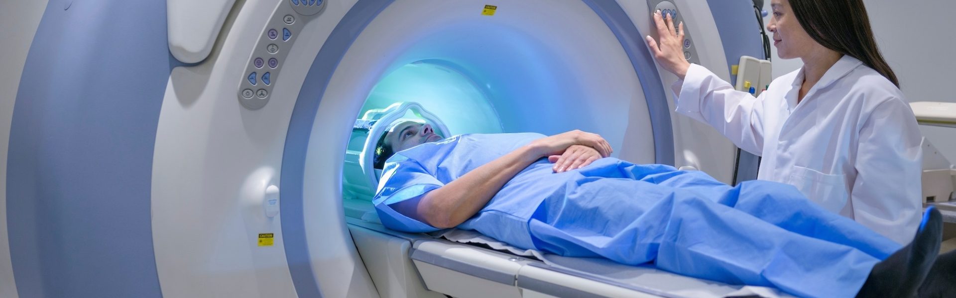
Virtual angiogram procedure
The virtual angiography examination of the coronary artery is performed under the constant supervision of health workers and doctors.
-
The patients communicate with the health workers through a microphone.
-
The health professionals hear the patient during the entire examination and is in constant contact with the patient.
-
In the first phase of the examination, a control image is taken before the intravenous medication is administered.
-
The images are obtained by “staining” the heart and coronary arteries with intravenously administered contrast.
-
When the drug is administered intravenously, the mouth may feel hot and/or metallic. This is a normal and temporary situation.
-
You will hear motor sounds when the imaging device starts up. During imaging, it is very important that the patient does not move, remains calm, and holds his breath. The breathing time is only one second.
Difference Between Conventional and Virtual Angiography
The following are the distinctions between normal angiography and virtual angiography, also known as tomographic angiography:
An artery is catheterized during a typical angiography operation, which is an invasive process. A catheter is passed through the artery, and contrast material is administered during this process. In contrast, virtual angiography is a non-invasive technique that involves injecting contrast material into a vein in the arm or wrist region without the need for a needle or catheter.
Routine angiography involves risks such as bleeding, infection, thrombosis, anesthesia, and vascular damage. However, virtual angiography is noninvasive, so there are fewer dangers.
A local anesthetic is used during a normal angiography treatment. In contrast, tomographic angiography often causes no discomfort, with the exception of a brief, faint warmth experienced after the contrast media injection. As a result, no local anesthesia is needed.
Normal angiography obtains more detailed pictures. In contrast, virtual angiography depends on the thickness of the vessel since it uses images taken from outside the vessel.
Heart and vascular disorders are diagnosed with normal angiography. All arteries besides the heart may be seen by exploring the virtual angiography near me option.
For the examination of stable coronary disease, a CT angiogram cost has proven to be more effective than invasive angiography in all risk groups. It has also been demonstrated that the use of CT angiography as a diagnostic test has increased significantly. This strengthens the case for increasing CT services and decreasing the number of patients listed for diagnostic angiography when there are no high-risk characteristics.